ABOUT

The scope of our research is broad, covering numerous areas including the cloning of genes involved in carcinogenesis, biological and structural analyses of proteins, analyses of animal models and clinical samples, and the development of new strategies for cancer prevention/diagnosis/therapy.

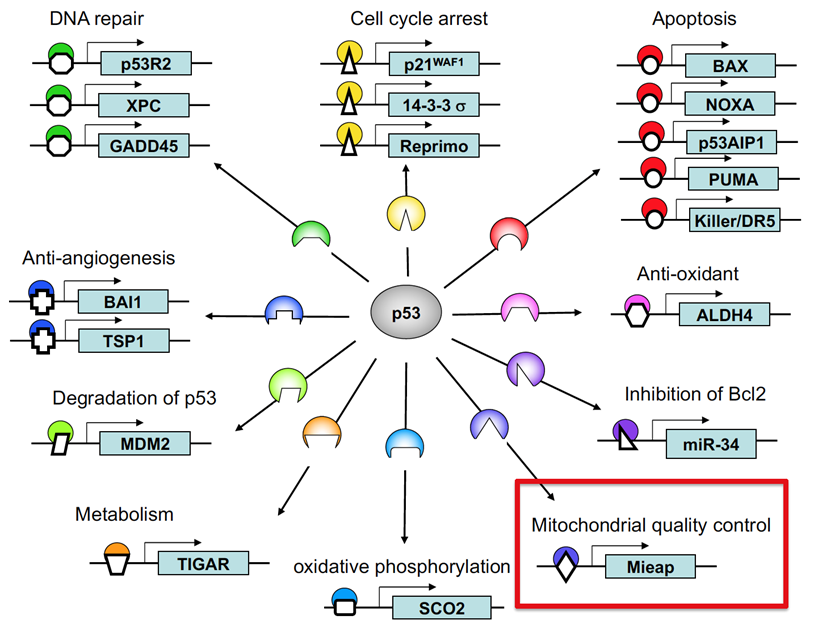
Hirofumi Arakawa (Chief, Division of Cancer Biology) has been devoted himself to the project of “Identification and characterization of p53 target genes”, and published a number of p53 target genes involved in apoptosis, cell cycle arrest, DNA repair, anti-angiogenesis, and others(Cell 2000, Nature 2000, Molecular Cell 2001, Nature Cell Biology 2003, Nature Genetics 2003, Nature Reviews Cancer 2004, Nature Genetics 2007, Cancer Research 2007)(Figure1).

Figure1. Tumor suppressor p53 regulates a large number of functions via transcriptional activation of its target genes as a transcription factor

p53/Mieap-regulated mitochondrial quality control


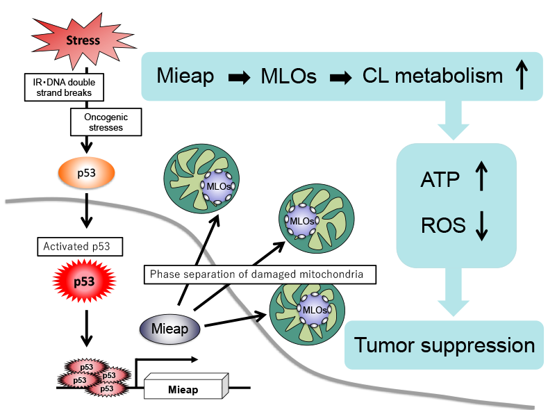
Mitochondria-eating protein (Mieap, also designated as SPATA18) was originally identified as a p53-inducible protein; its mRNA expression is directly regulated by the tumor suppressor p53 in response to various cellular stresses, including DNA damage (PLoS ONE 6: e16054, 2011). Mieap expression is lost in nearly 50% of human cancer cell lines due to promoter methylation. Mieap-deficient LS174T colorectal cancer cells exhibit increased mitochondrial reactive oxygen species (ROS) generation and decreased mitochondrial ATP synthesis. The mitochondrial ROS in Mieap-deficient colorectal cancer and gastric cancer cells enhance the migration and invasion activities of cancer cells under hypoxic conditions (Oncogenesis 5: e181, 2016; Scientific Reports 9: 2822, 2019). Mieap-deficient ApcMin/+ mice exhibit the promotion of intestinal tumor initiation and malignant transformation (Scientific Reports 5: 12472, 2015). Tumors in Mieap-deficient ApcMin/+ mice have revealed abnormal morphology of the mitochondria, such as enlarged size, round shape, and a decrease in the cristae structure. Recently, Mieap expression was reported to be defective in thyroid oncocytic cell tumors, which exhibit accumulation of abnormal mitochondria in tumor cells (Cancer Sci. 111: 2814–2823, 2020). Mieap-regulated mitochondrial quality control is inactivated in the tumor tissues of nearly 70% of colorectal cancer, 25% of breast cancer, 70% of gastric cancer, and 83% of esophageal cancer patients (Oncogenesis 5: e181, 2016; Cancer Sci. 109: 3910–3920, 2018; Biochem Biophys Res Commun. 529: 582-589, 2020). Mieap is associated with better survival in breast cancer patients (Am J Cancer Res. 11: 6060-6073, 2021). These observations suggest that Mieap plays a critical role in tumor suppression via its mitochondrial quality control function. However, the mechanism underlying the regulation of mitochondrial quality control by Mieap remains unclear.

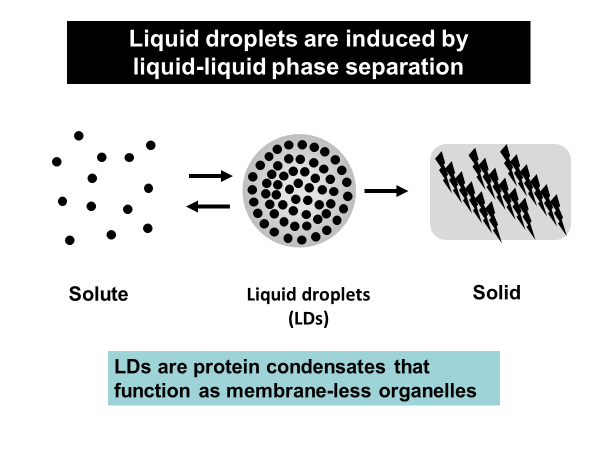
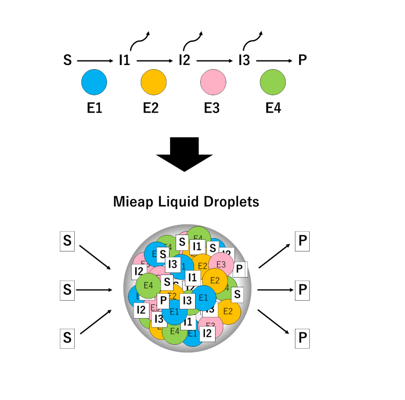
Liquid droplets function as membrane-less organelles in cells


Liquid droplets (LDs) in cells are protein condensates, which are composed of proteins, nucleic acids, and other macromolecular components (Droplets of life, 2022, Elsevier). LDs are formed by proteins with an intrinsically disordered region(s) (IDR) via liquid-liquid phase separation (LLPS) (Figure 2).

Importantly, LDs function as membraneless organelles that compartmentalize and facilitate cellular biological reactions. LDs are not surrounded by a lipid bilayer structure, which enables the dynamic exchange of reactants and products with their surroundings, facilitating biological reactions in cells. Furthermore, theoretically, LDs can appear and disappear anywhere within a cell in response to cellular stress and/or subcellular circumstances, exhibiting spatiotemporal dynamic properties. On the basis of these features, the concept of membraneless organelles is fundamentally different from the long-standing concept of membrane-bound organelles, such as the nucleus, lysosome, mitochondria, and endoplasmic reticulum, which stably exist and strictly segregate biomolecules and biological reactions in a lipid bilayer structure. It has been suggested that a number of membraneless organelles might exist within a cell, which could regulate various cellular biological reactions and activities, including transcriptional regulation, signal transduction, immunity, centrosome activity, and mitosis. This list is now rapidly growing.

Figure2. Liquid droplets are induced by liquid-liquid phase separation

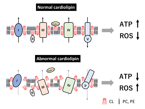
Cardiolipin (CL) is a unique and important phospholipid that is specific to mitochondria


CL is a phospholipid dimer, which has two phosphate residues and four fatty acyl chains. Because of this unique structure, it forms a cone shape, which plays a critical role in forming the curvature of the lipid membrane, contributing to maintenance of the mitochondrial cristae structure. CL is the only phospholipid that is specific to the mitochondria and is mainly located at the inner mitochondrial membrane and contact sites. CL interacts with many mitochondrial membrane proteins, including the electron transport chain complexes involved in oxidative phosphorylation and the ADP/ATP carrier; the interaction with CL promotes the activity of these proteins. CL stabilizes the structural assembly and activity of the respiratory super-complexes at the inner mitochondrial membrane. CL interacts with cytochrome c at the outer surface of the inner mitochondrial membrane, which plays a pivotal role in the life and death of cells by maintaining stable respiratory ATP production and inducing apoptosis, respectively. Therefore, altered CL metabolism and/or deterioration of CL quality causes various mitochondrial dysfunctions and diseases via abnormal cristae structures, decreased ATP synthesis activity, and increased ROS generation (Figure 3).

Figure3. Role of cardiolipin in mitochondrial proteins

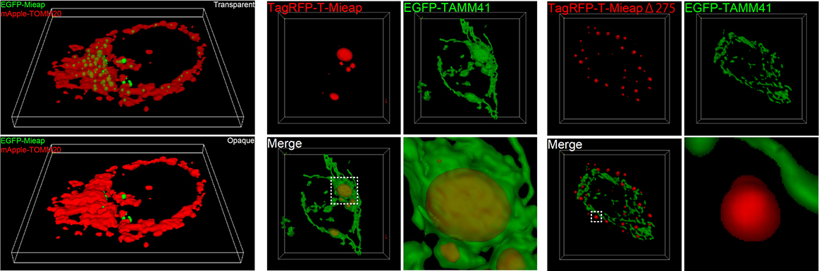
Mieap forms membraneless organelles to compartmentalize and facilitate cardiolipin metabolism


This year, we reported that Mieap is an IDR-containing protein that drives the formation of liquid droplets in the mitochondria (iScience 27: 108916, 2024). Mieap liquid droplets (MLDs) specifically phase separate the mitochondrial phospholipid cardiolipin. Lipidomic analysis of cardiolipin suggests that Mieap promotes enzymatic reactions involved in cardiolipin metabolism, including biosynthesis and remodeling. Accordingly, four cardiolipin biosynthesis enzymes, TAMM41, PGS1, PTPMT1, and CRLS1, and two remodeling enzymes, PLA2G6 and TAZ, are phase separated by MLDs. Mieap-deficient mice exhibited altered cristae structure in the liver and kidney mitochondria and a trend of obesity. These results suggest that Mieap drives the formation of membraneless organelles to compartmentalize and promotes cardiolipin metabolism at the inner mitochondrial membrane, thereby playing a possible role in mitochondrial quality control (Figure 4)

Figure4. Mieap liquid droplets function as membrane-less organelles to compartmentalize and facilitate cardiolipin metabolism

The Future


Autophagy and proteostasis are two major mechanisms in mitochondrial quality control. In addition to these, we suggest that the Mieap-regulated pathway is the third mechanism for mitochondrial quality control, in which Mieap maintains the integrity of mitochondria by regulating CL metabolism. Mieap maintains mitochondrial quality through homeostasis of the inner mitochondrial membrane by regulating CL metabolism, stabilizing oxidative phosphorylation, and suppressing mitochondrial ROS generation (Figure 5). Considering the role of Mieap in p53 function, we suggest that Mieap could act as a spatiotemporal and dynamic regulator/modulator in CL metabolism to suppress tumor initiation and progression.

Figure5. p53/Mieap suppresses tumors by regulating CL metabolism

References


  1. Ikari N, Honjo K, Sagami Y, Nakamura Y, Arakawa H. Mieap forms membrane-less organelles involved in cardiolipin metabolism. iScience. Vol.27: 108916. (2024)
    https://doi.org/10.1016/j.isci.2024.108916 [PubMed]
  2. Ikari N, Arakawa H. Identification of a mitochondrial targeting sequence in cathepsin D and its localization in mitochondria. Biochemical and Biophysical Research Communications. Vol.655: 25-34 (2023) [Times Cited: 1] [PubMed]
  3. Arakawa H. MLOs and control of metabolic pathways. Uversky V. Droplets of life. Elsevier. Chapter 14. 431-446. (2023)
    https://doi.org/10.1016/B978-0-12-823967-4.00023-3
  4. Hamada K, Kurashige T, Shimamura M, Arakawa H, Nakamura Y, Nagayama Y. MIEAP and ATG5 are tumor suppressors in a mouse model of BRAFV600E-positive thyroid cancer. Frontiers in Endocrinology. 13 : 932754. (2022) [PubMed]
  5. Futamura M, Tokumaru Y, Takabe K, Arakawa H, Asano Y, Mori R, Mase J, Nakakami A, Yoshida K. MIEAP, a p53-downstream gene, is associated with suppression of breast cancer cell proliferation and better survival. Am J Cancer Res. 11(12) : 6060-6073. (2021) [Times Cited: 3] [PubMed]
  6. Sano H, Futamura M, Gaowa S, Kamino H, Nakamura Y, Yamaguchi K, Tanaka Y, Yasufuku I, Nakakami A, Arakawa H, Yoshida K. p53/Mieap-regulated mitochondrial quality control plays an important role as a tumor suppressor in gastric and esophageal cancers. Biochemical and Biophysical Research Communications. 529 : 582-589. (2020) [Times Cited: 7] [PubMed]
  7. Gaowa S, Futamura M, Tsuneki M, Kamino H, Tajima JY, Mori R, Arakawa H, Yoshida K. Possible role of p53/Mieap-regulated mitochondrial quality control as a tumor suppressor in human breast cancer. Cancer Science. 109: 3910-3920 (2018) [Times Cited: 14] [PubMed]
  8. Nakamura Y and Arakawa H. Discovery of Mieap-regulated mitochondrial quality control as a new function of tumor suppressor p53. Cancer Science. 108: 809-817. (2017) [Times Cited: 28] [PubMed]
  9. Kamino H, Nakamura Y, Tsuneki M, Sano H, Miyamoto Y, Kitamura N, Futamura M, Taniguchi H, Kanai Y, Shida D, Kanemitsu Y, Moriya Y, Yoshida K, Arakawa H. Mieap-regulated mitochondrial quality control is frequently inactivated in human colorectal cancer. Oncogenesis. 5: e181 (2016). [Times Cited: 23][Pubmed]
  10. Tsuneki M, Nakamura Y, Kinjo T, Nakanishi R, Arakawa H. Mieap suppresses murine intestinal tumor via its mitochondrial quality control. Scientific Reports. 5: 12472 (2015). [Times Cited: 23][Pubmed]
  11. Miyamoto T, Kitamura N, Ono M, Nakamura Y, Yoshida M, Kamino H, Murai R, Yamada T, Arakawa H. Identification of 14-3-3gamma as a Mieap-interacting protein and its role in mitochondrial quality control. Scientific Reports. 2: 379 (2012). [Times Cited: 14][Pubmed]
  12. Nakamura Y, Kitamura N, Shinogi D, Yoshida M, Goda O, Murai R, Kamino H, Arakawa H. BNIP3 and NIX mediate Mieap-induced accumulation of lysosomal proteins within mitochondria. PLoS ONE. 7: e30767 (2012). [Times Cited: 48][Pubmed]
  13. Kitamura N, Nakamura Y, Miyamoto Y, Miyamoto T, Kabu K, Yoshida M, Futamura M, Ichinose S, Arakawa H. Mieap, a p53-inducible protein, controls mitochondrial quality by repairing or eliminating unhealthy mitochondria. PLoS ONE. 6: e16060 (2011). [Times Cited: 85][Pubmed]
  14. Miyamoto Y, Kitamura N, Nakamura Y, Futamura M, Miyamoto T, Yoshida M, Ono M, Ichinose S, Arakawa H. Possible existence of lysosome-like organella within mitochondria and its role in mitochondrial quality control. PLoS ONE. 6: e16054 (2011). [Times Cited: 72][Pubmed]
  15. Futamura M, Kamino H, Miyamoto Y, Kitamura N, Nakamura Y, Ohnishi S, Masuda Y, Arakawa H. Possible role of semaphorin 3F, a candidate tumor suppressor gene at 3p21.3, in p53-regulated tumor angiogenesis suppression. Cancer Research 67: 1451-1460 (2007). [Times Cited: 88][Pubmed]
  16. Bourdon A, Minai L, Serre V, Jais JP, Sarzi E, Aubert S, Chretien D, de Lonlay P, Paquis-Flucklinger V, Arakawa H, Nakamura Y, Munnich A, Rotig A. Mutation of RRM2B, encoding p53-controlled ribonucleotide reductase (p53R2), causes severe mitochondrial DNA depletion. Nature Genetics. 39: 776-780 (2007). [Times Cited: 465][Pubmed]
  17. Arakawa H. p53, apoptosis and axon-guidance molecules. Cell Death and Differentiation. 12: 1057-1065 (2005). [Times Cited: 65][Pubmed]
  18. Arakawa H. Netrin-1 and its receptors in tumorigenesis. Nature Reviews Cancer. 4: 978-987 (2004). [Times Cited: 234][Pubmed]
  19. Yoon KA, Nakamura Y, Arakawa H. Identification of ALDH4 as a p53-inducible gene and its protective role in cellular stresses. Journal of Human Genetics. 49: 134-140 (2004). [Times Cited: 207][Pubmed]
  20. Kimura T, Takeda S, Sagiya Y, Gotoh M, Nakamura Y, Arakawa H. Impaired function of p53R2 in Rrm2b-null mice causes severe renal failure through attenuation of dNTP pools. Nature Genetics. 34: 440-445 (2003). [Times Cited: 141][Pubmed]
  21. Tanikawa C, Matsuda K, Fukuda S, Nakamura Y, Arakawa H. p53RDL1 regualtes p53-dependent apoptosis. Nature Cell Biology. 5: 216-223 (2003). [Times Cited: 159][Pubmed]
  22. Ueda K, Arakawa H, Nakamura Y. Dual-specificity phosphatase 5 (DUSP5) as a direct transcriptional target for tumor suppressor p53. Oncogene 22: 5586-5591 (2003). [Times Cited: 113][Pubmed]
  23. Ng CC, Arakawa H, Fukuda S, Kondoh H, Nakamura Y. p53RFP, a p53-inducible RING-finger protein, regulates the stability of p21WAF1. Oncogene 22: 4449-4458 (2003). [Times Cited: 47][Pubmed]
  24. Iiizumi M, Arakawa H, Mori T, Ando A, Nakamura Y. Isolation of a novel gene, CABC1, encoding a mitochondrial protein that is highly homologous to yeast activity of bc1 complex. Cancer Research. 62: 1246-1250 (2002). [Times Cited: 48][Pubmed]
  25. Mori T, Anazawa Y, Iiizumi M, Fukuda S, Nakamura Y, Arakawa H. Identification of the interferon regulatory factor 5 gene (IRF-5) as a direct target for p53. Oncogene. 21: 2914-2918 (2002). [Times Cited: 138][Pubmed]
  26. Matsuda K, Yoshida K, Nakamura K, Nakamura Y, Arakawa H. p53AIP1 regulates the mitochondrial apoptotic pathway. Cancer Research. 62: 2883-2889 (2002). [Times Cited: 115][Pubmed]
  27. Yamaguchi T, Matsuda K, Sagiya Y, Iwadate M, Fujino AJ, Nakamura Y, Arakawa H. p53R2-dependent pathway for DNA synthesis in a p53-regulated cell cycle checkpoint. Cancer Research. 61: 8256-8262 (2001). [Times Cited: 143][Pubmed]
  28. Okamura S, Arakawa H, Tanaka T, Nakanishi H, Ng CC, Taya Y, Monden M, Nakamura Y. p53DINP1, a p53-Inducible Gene, Regulates p53-Dependent Apoptosis. Molecular Cell. 8: 85-94 (2001). [Times Cited: 320][Pubmed]
  29. Shiraishi K, Fukuda S, Mori T, Matsuda K, Yamaguchi T, Tanikawa C, Ogawa M, Nakamura Y, Arakawa H. Identification of fractalkine, a CXC3-type chemokine, as a direct target of p53. Cancer Research. 60: 3722-3726 (2000). [Times Cited: 58][Pubmed]
  30. Oda K, Arakawa H, Tanaka T, Matsuda K, Tanikawa C, Mori T, Nishimori H, Tamai K, Tokino T, Nakamura Y, Taya Y. p53AIP1, a potential mediator of p53-dependent apoptosis, and its regulation by Ser46-phosphorylated p53. Cell. 102: 849-862 (2000). [Times Cited: 1,135][Pubmed]
  31. Tanaka H, Arakawa H, Yamaguchi T, Shiraishi K, Fukuda S, Matsui K, Takei Y, Nakamura Y. A ribonucleotide reductase gene involved in a p53-dependent cell cycle checkpoint for DNA damage. Nature. 404: 42-49 (2000). [Times Cited: 839][Pubmed]